


Ways of infection by humans with helminths
- Biohelminthiasis (infection of animals).
- Transmissible helminths (transmitted from person to person).
- Geohelminthiasis (diseases caused by parasites that carry out one of their life cycles on earth).
- The way the parasite enters the body.
- The degree of adaptation of the helminth to the human body.
- Population density (number) of parasitic individuals?
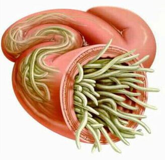
- The worm habitat (tissue parasites live in the soft tissue thickness and grooves live in the cavities of the hollow organs). Some helminths in different phases have both lumen and tissue. The stages of larvae and growing worms, as a rule, cause more pronounced pathological changes.
In the absence of recurrence, the number of adult parasites in the human body does not increase. This feature significantly distinguishes helminth infestations from diseases caused by bacteria, viruses, fungi and protozoa.
Worms in humans: symptoms
Helminthiasis is a disease characterized by 2 stages of the course (acute, from two weeks to two months) and chronic (from several months to several years).Symptoms of the acute phase of helminthiasis
The first signs of the disease may appear at different times (most often after 2-3 weeks, with exercise - after 2-3 days and with filariasis, the incubation period can last 6-18 months).
In the acute stage of the parasitic invasion, the most characteristic symptom is an allergic reaction (antibodies are produced to parasites of migratory larvae). Often in people infected with worms, itching occurs on the skin, prone to recurrent course, peripheral lymph nodes enlarged, generalized or local swelling, muscle aches and joint pain. Also, migratory pest larvae can cause chest pain, cough, choking, feces, nausea and vomiting.
At the same time, the acute phase of helminthiasis may be accompanied by more serious disorders (severe forms of pneumonia, hepatitis, allergic myocarditis, hepatosplenomegaly (enlarged liver and spleen), meningoencephalogram.The number of eosinophils in the blood increases (eosinophilia) and the normal quantity ratio between protein fractions is disturbed (dysproteinemia).
Signs of chronic helminthiasis
The symptoms of the chronic phase depend directly on which organ is "inhabited" by the parasites, as well as their size and number play an important role.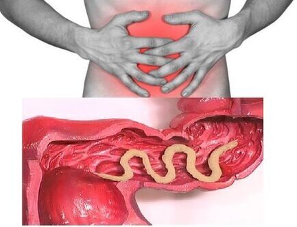 Thus, when parasitizing the intestines of individuals, the disease may be asymptomatic (except in cases of infection with very large parasites). The hallmarks of the chronic phase of intestinal helminthiasis are dyspeptic disorders. In children, the neurotic syndrome and pain syndrome is more intense. With massive infestation of worms, it is possible to develop intestinal obstruction, obstructive jaundice and pancreatitis.
By consuming all the substances necessary for their vital activity by the host body, the helminths cause digestive disorders, reduced absorption of vitamins, minerals, carbohydrates, proteins and fats. At the same time, worm waste inhibits the normal intestinal microflora and reduces the body's immune system.
In people with helminthiasis, due to a weakened immune system and increased cell division (as a result of the continuous repair of parasite-damaged tissue), the risk of malignancies increases significantly.
Types of helminth parasitization in the human body
The causative agents of human helminthiasis are 2 types of worms: round (nematode) and flat (tapeworms and worms).
Roundworms
Pinworm
The parasites that cause enterobiasis are small (up to 10 mm) worms with a thin cavity with a grayish white color. The infection occurs nutritionally (through the mouth). The reason for this is dirty hands. The eggs of the pest can be found in the soil, in the wool of infected animals, in unwashed vegetables and fruits, etc. At the same time, with enterobiasis, there are frequent cases of infection (especially in children), resulting from scratching the itching and the subsequent ingestion of eggs. 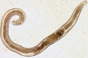 The cancer larva develops in the digestive system within two weeks. Having turned into an adult, the worm parasitizes the lower parts of the small and upper part of the large intestine.
Even in the larval stage, the pin begins to damage its host's body, producing enzymes that irritate the intestinal wall and lead to the development of an inflammatory process. Adult parasites attach to or penetrate the deeper layers of the intestinal mucosa, disrupting its integrity and contributing to the attachment of a secondary bacterial infection. In case of perforation of the worms in the wall of the small intestine, peritonitis may develop. Also, due to the irritation of the intestinal receptors, the motor and secretory functions of the gastrointestinal tract are disturbed, leading to the formation of gastroduodenitis, enteritis, etc. In childhood, long-term enterobiasis can cause nervous disorders and delayed physical development.
Ascaris
Ascaris is a large red-yellow spindle-shaped parasite that reaches 40 cm (females) and 15-25 cm (males) in adulthood. Without suction cups or other fasteners, the worm can move independently of food masses. Eggs produced by the female parasite are excreted in the feces.
Ascites infection occurs when mature eggs are swallowed with water or unwashed vegetables and fruits with soil particles. Once the eggs enter the intestines, mature larvae emerge from them. Then, penetrating the intestinal wall, they reach the heart through the bloodstream and from there enter the lungs. Through the lung cells, the worm larva enters the oral cavity again through the airway. After repeated ingestion, the parasite reaches the small intestine, where it develops into an adult. The worm lives for 12 months, then dies and is excreted in the feces. In the intestine of a host, both one and several hundred people can live.
In the intestinal phase of their existence, worms, endowed with the ability to spiral movements, can penetrate even the narrowest openings. This feature of the parasite often leads to the development of rather serious complications (obstructive jaundice or pancreatitis). Allergens secreted by worms can cause severe allergic reactions. Large numbers of adults can cause intestinal obstruction and worms entering the airway sometimes cause suffocation.
Vlasoglav
Vlasoglav, the causative agent of hair loss, is a white helminth parasite in the early part of the large intestine and grows to a size of 4-5 cm. The parasite feeds on blood and tissues of the rectal mucosa.The eggs of the female worm in the intestinal wall come out along with the feces. They grow in the environment (optimally in the soil). Eggs with parasite larvae mature enter the body through food, dirty hands, water or unwashed vegetables and fruits.
With small numbers of worms, the hair follicle is asymptomatic. In a severe stage (with massive invasion), the patient develops abdominal pain, develops severe diarrhea, sometimes accompanied by prolapse of the rectum. This condition is more common in debilitated children. With a moderate hair follicle phase, it is possible to delay a child's development.
Trichinella
The causative agent of trichinosis is a small round helminth 2-5 mm long. Infection occurs when you eat poorly cooked meat (pork, bear, wild boar).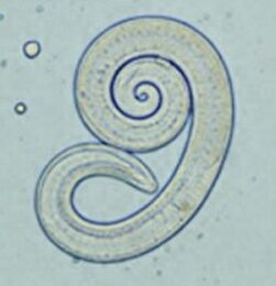 Penetrating into the intestines, the parasite larva matures in 3-4 days in the state of a sexually mature person. The lifespan of the worm is 40 days, after which the parasite dies. By piercing the intestinal wall, the larvae enter the bloodstream and are transported to all the organs of the human body, settling in the muscles. In this case, the respiratory and facial muscles, as well as the flexor muscles of the limbs, are most often affected.
In the first days after the invasion, patients complain of abdominal pain. Then, after about 2 weeks, the body temperature rises to 39-40 C, itching appears on the skin, muscle aches develop and the face swells. During this period, in the event of a mass infection, there is a significant risk of death. After about a month, the patient recovers. The parasite is enclosed in a spiral form, after which it dies within two years.
Hookworm and nekator
These two parasites are similar in both biological characteristics and disease-causing. In this regard, it is common to combine them with a common name (bracket). The worms, 10-15 mm long, parasitize at 12-p. intestine. It should be noted that this is one of the most common, but at the same time, parasites are rarely detected. Worm larvae enter the human body through the skin in contact with contaminated soil. Further, entering the bloodstream, like worms, they migrate to the lungs and then, through the bronchi, along with the expectorant sputum, to the digestive system. Ankylostoma parasitizes the intestine, attaching to the intestinal wall. The parasite, which feeds exclusively on blood, bites through the blood vessels that penetrate the mucous membrane, injecting an anticoagulant component there. On average, an adult can absorb 0. 05-0. 35 ml of blood per day. Therefore, the most characteristic symptom of this helminthiasis is iron deficiency anemia, as well as a change in the ratio of protein fractions (dysproteinemia).
Flatworms
Wide ribbon
This is one of the largest helminths, 10-20 meters long. The disease caused by this parasite is called bisexuality. The growth cycle of the worm begins with freshwater fish or crustaceans. The larva enters the human body, which is the ultimate owner of the broadband, along with the eggs or the infected fish fillets. Reaching the small intestine, the parasite attaches to its wall and grows into a mature individual within 20-25 days.
Bifurcation occurs in the context of digestive disorders and B12 deficiency anemia.Hepatic impairment
The parasite that causes opisthorchiasis is a flat worm 7-20 mm long. It should be noted that over 50% of cases of hepatic fluke infection (also called cat fluke) occur in Russians. The larvae of the parasite begin to develop after the eggs enter fresh water (from the snails that have swallowed them). They then penetrate the body of the fish (carp, carp, sea bream, cockroach). Human infection occurs when you eat contaminated fish meat that has not undergone adequate heat treatment. The hepatic larva from the small intestine penetrates the bile ducts and the gallbladder, securing there with the help of two suction cups.
In the acute phase of helminthiasis, the patient has pain in the upper abdomen, body temperature rises, nausea develops, muscle aches, diarrhea and skin rashes. The chronic course of retardation is manifested by symptoms of hepatitis, inflammation of the bile ducts, cholecystitis, disorders of the digestive system, nervous disorders, weakness and increased fatigue. The parasite leads to the development of irreversible changes and even after its expulsion, the patient does not suffer from chronic inflammatory processes and functional disorders.
Beef and pork ribbon
These parasites, almost identical in structure, are 5-6 meters long. Infection with teniarins and tenacity occurs due to the consumption of meat from cattle or pork that have been infected by the Finns (one of the intermediate forms of helminthiasis). Sustainable Finns, presented in the form of white bubbles measuring 0. 5 cm, attach to the wall of the human small intestine and turn into an adult in 3 months. The parasite of the film, which consists of more than 2000 parts, is constantly growing. In this case, the extremities, which contain eggs, break down and move independently along the colon into the anus, and then crawl out of the anus, or are released into the external environment along with the stool. The most characteristic symptoms of helminthiasis are a disorder of the digestive tract.
Echinococcus
For this parasite, a person is an intermediate host. The worm parasitizes the human body in the form of Finns. The final owner of the echinococcus is a wolf, dog or cat.  Infection occurs through food contact with animals and environmental objects sown with Echinococcus eggs. Once they enter the intestine, they grow from these ossicles (six hooks). From the intestines, they enter the bloodstream and are transported throughout the body.
The "favorite" parasitic sites of the worm are the liver and lungs. By installing these organs, the larva transforms into a Finnish (echinococcal cyst), which, gradually increasing in size, begins to destroy nearby tissues. Often, echinococcosis in the diagnostic process is perceived as a tumor of benign or malignant origin. In addition to mechanical impact (compression of organs and blood vessels), rupture of the echinococcal bladder sometimes occurs. This condition can cause toxic shock or the formation of multiple new cysts.
Alveococcus
This parasite, considered a type of echinococcus, is the cause of one of the most dangerous helminths, which is similar in severity to cirrhosis and liver cancer. Infection occurs when the osphere (eggs with mature larvae) enters the intestine. There, the fetus leaves the egg and, penetrating the intestinal wall, enters the bloodstream. In addition, with the flow of blood, the parasite spreads to all tissues and organs of the body (most often located in the liver). This is where the main developmental stage in the larvae begins (a multi-chamber bubble is formed, a laryngeal cyst is formed). Each chamber contains the embryonic head of the parasite, which continues to grow gradually. Laurocysts are very aggressive formations that grow continuously due to swollen blisters and also have the ability to grow in the liver, such as cancerous metastases. Necrotic changes due to disturbances in the function of blood vessels There are necrotic changes in nearby tissues. Scattering in nearby structures, the cell forms fibrous nodules with polymer bubble inclusions. This condition can last for several years and therefore requires mandatory surgery.
Diagnostics of helminthiasis
Diagnoses of helminth infestations include the following activities:
- A thorough history taking to help discover the possible causes of the infection.
- laboratory tests of feces, blood, intestinal contents 12p, rectal and anal mucus, muscle tissue, pulmonary sputum, bile. The analysis may reveal eggs, fragments or fragments of parasites. At the same time, an increased content of eosinophils in the blood is also an indication of the presence of helminthiasis.
- in the diagnosis of diseases caused by larval stages or tissue parasites, serological studies are performed (ELISA, RSK, indirect welding reaction, immunofluorescence analysis, etc. ).
- Ultrasound, CT and endoscopic examinations are prescribed to detect helminths that affect the liver tissue.
Human worms: cure
In the acute phase of a parasitic infection, the patient is prescribed detoxification and desensitization treatment. In severe cases of the disease (liver fragments, trichinosis), glucocorticoids are used according to medical indications.
Special anthelmintic chemotherapeutic agents are prescribed as special treatment drugs, taking into account the nature of the pathogen.
At the same time, it is recommended that the patient take antihistamines and enterosorbents. The final stage of treatment involves the use of probiotics that normalize the intestinal microflora.A special diet is also recommended (food should be digestible and low in fat).
During the period of anthelmintic treatment, the patient is obliged to strictly observe personal hygiene (in order to avoid recurrence). At the same time, for many helminths, all family members and those who are in constant contact with the infected must be treated.
Prevention of helminthiasis
- Maintain personal and public hygiene.
- Strict adherence to cooking technology.
- Regular examination and preventive treatment of pets.
- Clean washing of fresh vegetables, fruits and herbs.
- The proper handling of river fish?
- Avoid eating raw, lightly salted and dried fish.